
Understanding symptoms, diagnostic procedures, and long-term recovery pathways for peripheral nerve trauma following canine puncture wounds.
Mechanisms of Canine-Induced Nerve Trauma
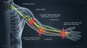
Peripheral nerves act as the complex electrical wiring of the human body, sending sensory feedback to the brain and transmitting motor commands to muscles. Canine teeth function as sharp wedges paired with massive clamping pressure, damaging nerve trunks through three primary mechanical pathways:
- Neurapraxia (Nerve Compression): Bluntt force or heavy compression crushes the nerve sheath without severing underlying axons. Signal conduction is temporarily blocked, but the structural continuity remains intact.
- Axonotmesis (Axonal Disruption): Sharp tearing or stretching forces disrupt internal nerve fibers (axons) while preserving the surrounding connective tissue sheaths. Regeneration is possible but prolonged.
- Neurotmesis (Complete Severance): Deep puncture wounds or avulsion injuries cut directly through both the axons and encapsulating nerve tissue, causing total disruption of sensory and motor signals.
Recognizing Symptoms of Peripheral Nerve Loss
In the immediate hours after an attack, local swelling and high systemic adrenaline levels can mask nerve damage. As acute inflammation stabilizes, victims should monitor for distinct neurological symptoms across affected limbs:
- Sensory Deficits: Persistent numbness, localized loss of temperature perception, or chronic tingling (“pins and needles”) distal to the bite location.
- Motor Dysfunctions: Sudden inability to extend fingers, reduced grip strength, foot drop, or difficulty performing routine fine-motor tasks.
- Neuropathic Pain: Intense, burning sensations or sudden electric shock-like pains triggered by light touch or temperature changes.
- Trophic Changes: Long-term changes in skin texture, reduced sweating in localized areas, or visible muscle wasting (atrophy).
Diagnostic Protocols and Medical Evaluations

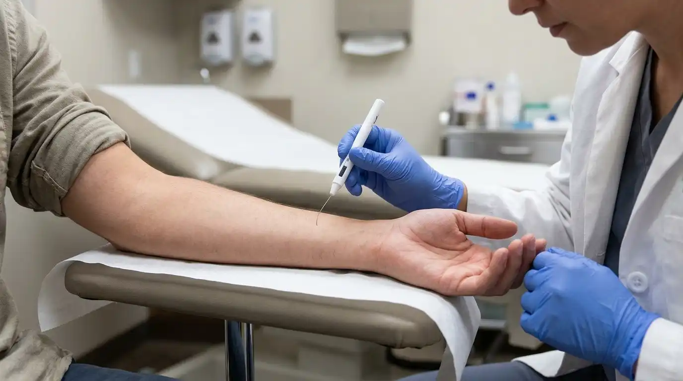
Accurate assessment of nerve trauma requires specialized neurological testing beyond basic physical examination. Emergency room staff and orthopedic specialists utilize structured diagnostic pathways to map out the location and severity of structural damage:
- Two-Point Discrimination Testing: Measures sensory perception boundaries across cutaneous skin regions to detect localized loss of touch sensitivity.
- Electromyography (EMG): Evaluates electrical activity within specific muscle groups at rest and during contraction to check for nerve signal disruption.
- Nerve Conduction Studies (NCS): Tracks the speed and signal strength of electrical impulses traveling across peripheral nerve segments.
- High-Resolution Ultrasound & MRI: Provides non-invasive visual imaging of soft tissue structures to identify nerve neuromas, scarring, or complete continuity gaps.
Treatment Pathways: Surgical vs. Conservative Management
Treatment strategy depends directly on injury severity, anatomical location, and whether the nerve sheath remains continuous:
Non-Surgical Management
Mild neurapraxic injuries often recover spontaneously over weeks or months. Patients are managed with temporary splinting to prevent joint contractures, target pain management using neuropathic medications, and structured physical therapy to maintain range of motion while nerve pathways recover.
Microsurgical Intervention
When nerve continuity is lost (neurotmesis) or severe scarring prevents regeneration, neurosurgical repair becomes necessary. Surgeons employ advanced techniques such as primary neurorrhaphy (direct end-to-end microscopic suturing), autologous nerve grafting, or synthetic conduit placement to bridge structural gaps.
Rehabilitation, Recovery Timelines, and Long-Term Outlook
Regeneration of damaged peripheral nerves is a slow process, typically progressing at a rate of 1 millimeter per day (roughly one inch per month). Full functional recovery can require anywhere from six months to over two years, depending on the distance between the injury site and the target muscles.
Consistent occupational therapy, functional electrical stimulation (FES), and dedicated home exercise regimens are essential during this extended window to keep target muscles responsive. For official guidelines on post-injury infection prevention, wound monitoring, and public safety data, refer to resources from the Centers for Disease Control and Prevention.